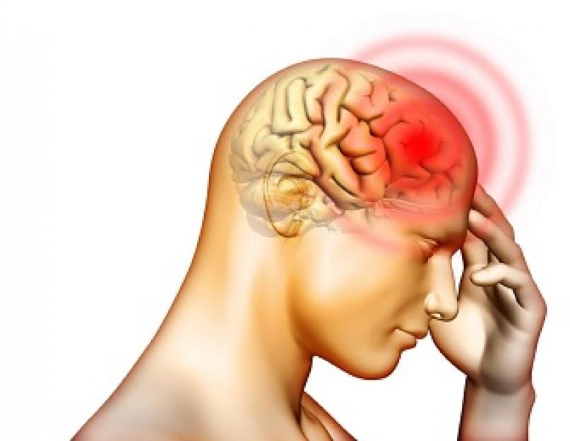
Ischemic stroke is a serious disease in which blood supply to a certain area of the brain is cut off. The disease occurs in people aged 50 years and older.
Ischemic stroke in the territory of the right middle cerebral artery is fatal in 1/3 of cases.
Causes and symptoms
Right-sided ischemic stroke occurs due to:
The following symptoms indicate a right-sided stroke:

If you notice any of these signs, call an ambulance immediately.
The consequences depend on the severity of the brain damage, the correct treatment and recovery procedures. In old age, these factors play a decisive role. Timely first aid provides hope for a favorable prognosis for life.
 The time factor is of great importance, because up to 25% of patients diagnosed with right-sided ischemic stroke die in the first 4 weeks.
In almost all cases, the cause of death is cerebral edema and pathologically caused dislocation of its structures. Most deaths occur in the first 48 hours after the onset of a stroke.
In 70% of surviving patients, disabling neurological disorders are noted. Six months after the hemorrhage, neurological disorders are observed in 40% of patients, and by the end of 12 months in 30%. If body functions have not been restored within a year, this condition will remain forever.
Based on the clinical picture of the disease, specialists make a prognosis for the patient’s life and health:

In the first 3 months after a stroke, the health prognosis is as follows:
- Motor functions begin to gradually improve. In this case, the lower limb recovers faster, unlike the arm. If by the end of the first month arm movement has not resumed, this is a bad sign.
- The swallowing reflex is restored.
- The intellectual components are gradually being improved.
During the first 5 years after suffering a right-sided stroke, the prognosis for the patient’s life and health is unfavorable with the following accompanying factors:

In this situation, 30% of patients developed a recurrent ischemic stroke.
Diagnosis and treatment
Only a specialist, based on the symptoms, age of the patient and concomitant diseases, selects effective methods examinations.
Main research:

The neurologist may prescribe additional studies if necessary.
Therapy in a hospital setting
Therapy for right-sided cerebral infarction in a hospital setting consists of basic and specific treatment.
The treatment strategy is aimed at reducing mortality, minimizing neurological deficits and ensuring a favorable prognosis for the patient's life.
Basic treatment includes:

Specific treatment is necessary to eliminate the causes of the pathology:

When treating ischemic hemorrhage in the right hemisphere of the brain, it is important to correctly and timely influence all parts of the pathogenesis.
Rehabilitation and prevention
The goal of restorative procedures is to stabilize neurological symptoms and stimulate their regression. At the cellular level, new contacts are formed between neurons, and processes of excitability and conductivity are activated.
Medicines stimulate the metabolism of neurons, these include:

Work to restore the patient's ability to work begins immediately after his transfer to the general ward. Rehabilitation at this time consists of an individual program of massage, physical therapy and electrical stimulation. Thanks to massage and exercise therapy, the patient will begin to restore sensitivity in the limbs, and connections between neurons will be activated.
It is worth noting that massotherapy at the stage of early recovery consists of light stroking, which helps to increase muscle tone.
Features of the recovery phase:

Measures to prevent cerebral infarction include:

People at risk are:
- WITH chronic diseases hearts.
- Having diabetes.
- Suffering from high blood pressure and hypercholesterolemia.
Catad_tema Stroke - articles
Ischemic stroke: malignant infarction in the middle cerebral artery. Clinical recommendations.
Ischemic stroke: malignant infarction in the middle cerebral artery territory
ICD 10: I63.0, I63.1, I63.2, I63.3, I63.4, I63.5, I63.8
Year of approval (revision frequency): 2016 (revised every 10 years)
ID: KR573
Professional associations:
- Association of Neurosurgeons of Russia
Approved
Agreed
2. Dzhindzhikhadze R.S., Dreval, ON, Lazarev V.A. Decompressive craniectomy for intracranial hypertension. – M.: GEOTAR-Media, 2014.
3. Krylov V.V., Nikitin A.S., Dashyan V.G., Burov S.A., Petrikov S.S., Asratyan S.A. Surgery of massive ischemic stroke. – M.: GEOTAR-Media, 2016.
4. Krylov V.V., Petrikov S.S., Belkin A.A. Lectures on neuroreanimation. – M.: Medicine, 2009.
5. Lebedev V.V., Krylov V.V., Tkachev V.V. Decompressive craniotomy. Neurosurgery 1998; 2:38-43.
6. Nikitin A.S., Asratyan S.A. Functional outcome after decompressive craniotomy in patients with massive malignant ischemic stroke. Neurological Journal 2016; 3(21): 142-145.
7. Nikitin A.S., Krylov V.V., Burov S.A., Petrikov S.S., Asratyan S.A., Kamchatnov P.R., Kemezh Yu.V., Belkov M.V., Zavalishin E.E. Dislocation syndrome in patients with a malignant course of massive ischemic stroke. Journal of Neurology and Psychiatry named after S.S. Korsakov 2015; 3 Special issue “Stroke”: 20-26.
8. Shevelev O.A., Tardov M.V., Kalenova I.E., Sharinova I.A., Shmyrev V.I. Craniocerebral hypothermia in the acute period of ischemic stroke: changes in the degree of neurological deficit and features of cerebral blood flow. Kremlin Medicine. Clinical Journal 2012;3: 34-36.
9. Bereczki D. Mannitol for acute stroke. Cochrane Database Syst. Rev. 2007; 3: CD001153
10. Christensen M. Cerebral apoplexy (stroke) treated with or without prolonged artificial hyperventilation. Cerebral circulation, clinical course, and cause of death. Stroke 1973; 4: 568-619.
11. Dohmen C. Identification and clinical impact of impaired cerebrovascular autoregulation in patients with malignant middle cerebral artery infarction. Stroke 2007; 38: 56-61.
13. Hacke W. "Malignant" middle cerebral artery territory infarction: clinical course and prognostic signs. Arch. neurol 1996; 53:309-315.
14. Krieger D. Cooling for acute ischemic brain damage (COOL AID): an open pilot study of induced hypothermia in acute ischemic stroke. Stroke 2001; 32:1847-1854.
15. Quizilbash N, Lewington SL, Lopez-arietta J. Corticosteroids for acute ischemic stroke. Cochrane library. Oxford (United Kingdom): Update software.- 2001 (1).
16. Qureshi A.I., Suarez J., Yahia A.M. et al. Timing of neurological deterioration in massive middle cerebral artery infarction: a multicenter review. Crit. Care Med 2003; 31: 272-277.
17. Schwab S., Schwarz S., Spranger M. Moderate hypothermia in the treatment of patients with severe middle cerebral artery infarction. Stroke 1998; 29(12): 2461-2466.
18. Simard D., Paulson O. Artificial hyperventilation in stroke. Trans. Am. Neurol. Assoc. 1973; 98: 309-310.
19. Steiner T., Pilz J., Schellinger P. Multimodal online monitoring in middle cerebral artery territory stroke. Stroke 2001; 32 (11): 2500-2506.
20. Wijdicks E., Diringer M. Middle cerebral artery territory infarction and early brain swelling: progression and effect of age on outcome. Mayo Clin. Proc 1998;73(9):829-836.
21. Wijdicks E., Sheth K., Carter B.
et al. Recommendations for the management of cerebral and cerebellar infarction with swelling: a statement for healthcare professionals from the American Heart Association/American Stroke Association. Stroke 2014; 45(4): 1222-1238.
22. Woodcock J., Ropper A., Kennedy S. High dose barbiturates in non-traumatic brain swelling: ICP reduction and effect on outcome. Stroke 1982;.13: 785-787.
|
Appendix A1. Composition of the working group |
Krylov Vladimir Viktorovich Academician of the Russian Academy of Sciences, Director of the Clinical medical center |
|
Moscow State Medical and Dental University named after. A.I. Evdokimova, head of the department of emergency neurosurgery at the Research Institute of Emergency Medicine named after. N.V. Sklifosovsky, Head of the Department of Neurosurgery and Neuroresuscitation, Moscow State Medical and Dental University. A.I. Evdokimova |
Dreval Oleg Nikolaevich |
|
Doctor of Medical Sciences, Professor, Head of the Department of Neurosurgery of the Russian Academy of Postgraduate Education of the Ministry of Health of the Russian Federation |
Dzhindzhikhadze Revaz Semenovich |
|
Candidate of Medical Sciences, Associate Professor of the Department of Neurosurgery of the Russian Academy of Postgraduate Education of the Ministry of Health of the Russian Federation |
Lazarev Valery Alexandrovich |
|
Doctor of Medical Sciences, Professor of the Department of Neurosurgery of the Russian Academy of Postgraduate Education of the Ministry of Health of the Russian Federation |
Dashyan Vladimir Grigorievich |
|
Doctor of Medical Sciences, Professor of the Department of Neurosurgery and Neuroresuscitation, Moscow State Medical and Dental University. A.I. Evdokimova |
Nikitin Andrey Sergeevich |
|
Candidate of Medical Sciences, assistant at the Department of Neurosurgery and Neuroresuscitation, Moscow State Medical and Dental University. A.I. Evdokimova |
Petrikov Sergey Sergeevich |
- Doctor of Medical Sciences, Professor of the Russian Academy of Sciences, Deputy Director of the Research Institute of Emergency Medicine named after. N.V. Sklifosovsky, professor of the department of neurosurgery and neuroreanimation of the Moscow State Medical and Dental University named after. A.I. Evdokimova
- Neurosurgery
- Neurology
Table P1. Levels of evidence, indicating the classification of levels of evidence used
Table P2. Levels of strength of evidence indicating the classification of levels of strength of evidence used
Appendix B. Patient management algorithms
Algorithm 1. Stroke duration less than 24 hours
no Yes
|
Decompressive craniotomy (in the absence of contraindications) |
Appendix B: Patient Information
Patients with a malignant infarction in the middle cerebral artery in the acute period of the disease are deeply disabled. After discharge from the hospital, where the patient was treated for stroke, comprehensive rehabilitation in a specialized center is indicated, aimed at partial regression of the neurological deficit. The nature of rehabilitation and the number of courses is determined by a rehabilitation specialist. Outside the rehabilitation center on an outpatient basis, the patient is under the supervision of a neurologist at the place of residence, who determines treatment. Activities aimed at regressing neurological deficits (physical therapy, massage, classes with a speech therapist), prevention and treatment of extracranial complications continue. All patients require care in the first 3-6 months after a stroke. During rehabilitation, 3-6 months after a stroke, 50% of patients are restored to the level of moderate disability with the ability to walk independently and self-care.
PACIFIC STATE MEDICAL UNIVERSITY
Department of Psychiatry and Neurology
Head Department: Doctor of Medical Sciences, Professor Ulyanov I.G.
Teacher: Doctor of Medical Sciences, Professor Gulyaeva S. E.
DISEASE HISTORY
Clinical diagnosis
Related: Stage III hypertension
Completed by: student 402 gr. l/f
Barabash A.S.
Vladivostok
PASSPORT PART
Age: 48 years old
Nationality Russian
marital status: Not maried
Profession: driver
Location:
Date of admission to the clinic: 03/29/2015
COMPLAINTS
Weakness in the left hand and its numbness, as well as speech impairment.
ANAMNESlS MORBI:
On the evening of March 29, I began to feel numbness in my left arm, it became weak. Then he called a friend and noted that he could not express himself clearly, his speech was impaired. Then he called an ambulance medical care, who took him to the hospital of the Regional Clinical Hospital No. 1. The emergency doctor stated that the blood pressure was 260/120 mm. rt. st
ANAMNESIS VITAE:
Denies viral hepatitis, tuberculosis, sexually transmitted diseases and AIDS. There were no injuries, surgeries or head injuries. There is no allergic history. Increased blood pressure from age 35. Material and living conditions at various periods of life are satisfactory. Family history: the patient’s mother had hypertension and obesity. Bad habits: does not smoke. Denies alcohol abuse and drug use. There are no occupational hazards.
STATUS PRAESENS:
The condition is of moderate severity. Consciousness is clear. Body temperature is normal (36.6). He understands spoken speech. Has 4 degrees of obesity. Height 173 cm, weight 199 kg.
The skin, visible mucous membranes are pink, have normal moisture; subcutaneous fat tissue is overdeveloped.
The occipital, cervical, parotid, anterior cervical, submandibular, supraclavicular, subclavian, axillary, inguinal and popliteal lymph nodes are not palpable.
Mild swelling is detected in the ankle joint. The degree of muscle development and their tone are normal. There is no trembling or tremor of individual muscles. Deformation of the bones and changes in the terminal phalanges of the fingers and toes are not detected. The configuration of the joints is normal, skin color and local temperatures in the joint area are also normal. There is no curvature of the spine.
Respiratory organs: The chest is hypersthenic. Type of breathing - mixed, respiratory rate - 21 per minute, breathing through the nose is free; silent, rhythmic, moderate depth. On palpation, the chest is painless, the right and left halves evenly participate in the act of breathing. The lower limit of the lungs is within normal limits. Percussion - clear pulmonary sound. Auscultation - vesicular breathing, no wheezing.
CVS organs:
Limits of relative dullness of the heart:
Left: in the 5th intercostal space on the midclavicular line
Right: in the 4th intercostal space 1 cm outward from the right edge of the sternum
Upper: on the 3rd rib, along the left parasternal line.
The heart configuration is normal. The width of the vascular bundle in the second intercostal space is 7 cm. Auscultation: heart sounds are clear, pure, rhythmic, accent of the second tone at the point of auscultation of the aortic valve. HR-95. Noises and pathological rhythms are not heard. No splitting or splitting of tones was detected. There is no pericardial friction rub. A/D 140/90 mm. rt. Art.
Digestive organs: The tongue is moist and clean. The mucous membrane of the inner surface of the lips, cheeks, and palate is pink and clean. The tonsils are not enlarged. Stomach correct form, normal size, soft and painless on palpation. Evenly participates in the act of breathing. There is no visible peristalsis of the stomach and intestines. The pancreas is painless on palpation, liver dimensions according to Kurlov:
)10cm )9cm )8 cm The lower edge of the liver is at the level of the costal arch, rounded, soft, painless; the gallbladder is not palpable. The Shchetkin-Blumberg phenomenon is negative. Organs of the genitourinary system: Upon examination, the lumbar region is not changed, the kidneys are not palpable; the tingling symptom is negative. Kidneys, bladder not palpable. There are no dysuric disorders. Primary and secondary sexual characteristics are formed according to gender and age. There are no growth disturbances. The thyroid gland is not visible and not palpable. NEUROLOGICAL STATUS Consciousness is clear. The patient is oriented in time, place and space. Dysarthria, with increased speech activity. Meningeal symptoms: Kernig's symptoms are negative, upper, middle and lower Brudzinski's symptoms are negative. There is no stiffness of the neck muscles. Bechterew's syndrome and Gordon's syndrome are absent. Cranial nerves. I pair - olfactory nerves. Smells are distinguished and differentiated from both nostrils. II pair - optic nerve. No decrease in visual acuity is noted. Color discrimination is not impaired. There is no loss of visual fields. The fundus was not examined. ΙΙΙ, ΙV, VI pairs - oculomotor, trochlear, abducens nerves. The palpebral fissures are symmetrical. The movements of the eyeballs are not limited in volume. The pupils are identical, of a regular round shape. The reaction of the pupils to light is direct and friendly. The reaction to convergence is well expressed. The pair is the trigeminal nerve. Palpation of the trigeminal points is painless. Movement of the lower jaw is not limited. The tone of the masticatory and temporal muscles is the same. Corneal and conjunctival reflexes are alive, identical on both sides. II pair - facial nerve. The face at rest is asymmetrical, there is a drooping of the left corner of the mouth. The patient may close his eyes and frown his eyebrows, wrinkle his forehead, and bare his teeth (symmetrically). There is no lacrimation or dry eyes observed in the vestibulocochlear nerve. The hearing is not impaired, he understands whispered speech from 6 meters. Nystagmus is not observed., X pairs - glossopharyngeal and vagus nerves. Swallowing and phonation were preserved. The soft palate is mobile. Palatine and pharyngeal reflexes are alive on both sides. The pair is an accessory nerve. There are no muscle atrophies or deformities of the sternocleidomastoid muscle. Head turns are preserved. XII pair - hypoglossal nerve. Speech is not clear, the tongue deviates to the left. There are no atrophies or fibrillary twitches. Motor sphere When examining the muscles of the limbs and trunk, muscle atrophy is not determined, fibrillary and fascicular twitching is not detected. Movements of the upper limbs are possible in full: · in the shoulder joint, movements are performed in the frontal plane - abduction up to 90 degrees and around the long axis of the shoulder - rotation inward and outward 20 degrees. In the sagittal plane - flexion up to 130, extension up to 35 degrees. The arm extended forward to a horizontal position can be retracted back to an angle of 120 degrees and brought towards the opposite arm (towards the midline of the body) to an angle of 30 degrees. · At the elbow joint, the forearm is flexed to an angle of 140 degrees. · In the wrist joint, movements are made towards the palmar surface - palmar flexion of the hand up to 50 degrees, towards the rear - dorsiflexion (or dorsal extension) up to 50 degrees, deviation of the hand to the radial side (abduction) - 15 degrees and ulnar (adduction) - 35 degrees . Prosupination movements of the hand (turning inward and outward) together with the forearm are performed within 80 degrees in both directions. No gear, jackknife, or plastic hypertonicity phenomena were detected. Strength in the muscles of the shoulder, forearm, hand and fingers of the right hand is 5 points, of the left hand - 4 points. Movements of the lower extremities are possible in full: · IN hip joint flexion-extension movements are performed from the sagittal plane: flexion up to 120 degrees, extension up to 10 degrees. In the frontal plane, abduction up to 30 degrees and adduction up to 30 degrees are performed. Rotational movements are determined in the position of full extension of the hip or when it is flexed at the hip joint at an angle of 90 degrees. · The range of these movements occurs within 45 degrees in one direction (internal rotation) and the other (external rotation). Further movements in the hip joint are possible, but they are performed with the pelvis. · In the ankle joint: plantar flexion up to 45 degrees, dorsiflexion (extension) up to 25 degrees. Adduction and abduction of the forefoot within 30 degrees, carried out through movement in small joints. The strength of the muscles of the left thigh, lower leg and foot is 4 points, the right thigh, lower leg and foot is 5 points. The pace of movement is sufficient. Bare's test: upper and lower are positive on the left. Reflex sphere Deep reflexes from hands: flexion-elbow (C 5-C6) - yes, alive, stronger on the left wrist (C 5-C8) - present, alive, stronger on the left extensor elbow - yes, alive, stronger on the left upper (D 7-D8) - yes, lowered on the left middle (D9 - D10) - yes, reduced on the left lower (D11-D12) - yes, lowered on the left Deep reflexes from the legs: knee (L 3- L4) - yes, alive, stronger on the left Achilles (L5 - S1) - yes, alive, stronger on the left Pathological reflexes of oral automatism are absent. Pathological foot reflexes: Babinski's sign(with line irritation of the sole, reflex extension of the fingers) negative Rossolimo's symptom(reflex flexion of fingers II - V as a result of a short blow to their tips with a hammer) negative Bekhterev-Mendel's sign(flexion of the II - V fingers when tapping with a hammer on the anterior outer surface of the dorsum of the foot) negative Zhukovsky's symptom(plantar flexion of fingers II - V when tapping with a hammer on the sole under the fingers) negative. Oppenheim's sign(as a result of pressing with a pad thumb along the anterior surface of the tibia from top to bottom there is a reflex extension of the thumb) negative. Gordon's sign(as a result of hand compression of the mass of the calf muscle, a reflex extension of the thumb is observed) negative. Poussep's sign(abduction of the fifth finger with line irritation of the outer edge of the foot), negative. Coordination area Gait is not impaired. Static tests: Romberg position - the patient is stable. Dynamic tests: Finger-nose test: performs correctly. Heel-knee test: performed correctly Sensitive Sphere Hypoesthesia in left limbs. Functions of the pelvic organs The function is not affected. Higher cortical functions Cognitive functions preserved DATA OF ADDITIONAL RESEARCH METHODS 1.General urine analysis: red blood cells +++ 250 bilirubin- urea+ 16 protein ++ 1g density 1.025 leukocytes + 25 2.Urinalysis according to Nechiporenko: leukocytes - 18,000, erythrocytes - 82,000. 3.No helminth eggs were found. Albumin 46.8 g/l Total protein 81g/l Cholesterol 6.8 mmol/l Triglycerides 1.44 mmol/l Urea 6.8 mmol/l Total bilirubin 10.3 µmol/l Direct bilirubin 3.4 µmol/l In PSMA there is increased peripheral resistance. CPP on the right SMA is 80 mm. rt. Art., left SMA-106 mm. rt. Art. AD-198/119 mm. rt. Art. SYNDROME DIAGNOSIS 1. Central paresis VII and XII pairs ChMN on the left: · Drooping of the left corner of the mouth · Dysarthria · Tongue deviation to the left Central left hemiparesis · Strength in the muscles of the shoulder, forearm, hand and fingers of the left hand - 4 points. The strength of the muscles of the left thigh, lower leg and foot is 4 points. · Deep reflexes from the hands are preserved S>D · Abdominal: upper, middle, lower - reduced S>D · Deep reflexes from the legs, knees, Achilles - preserved S>D Sensory impairment in the form of hypoesthesia in the left extremities. Movement disorders in the form of central left-sided hemiparesis indicate damage to the pyramidal tract, which begins in the right hemisphere in the neurons of the anterior central gyrus, then it goes to the internal capsule (anterior two-thirds of the posterior thigh), then it passes through the middle part of the cerebral peduncles, descending through the base of the pons and in the lower part of the medulla oblongata passes to the opposite side and approaches the anterior horns. Central paresis of the VII and XII pairs of the cranial nerves indicates a unilateral lesion of the corticonuclear pathway passing in the knee of the internal capsule, in the middle part of the cerebral peduncles. The path crosses when approaching the cores. Sensory disorders in the form of left-sided hemihypesthesia. Pathways of superficial sensitivity (pain, temperature and partially tactile). The first neurons for all types of sensitivity lie in the spinal ganglia. The fibers from them, through the dorsal roots, enter the dorsal horns of the spinal cord of the same side, where the second neuron is located, then the fibers pass through the anterior commissure to the opposite side, obliquely rising 2-3 segments higher, and as part of the anterior sections of the lateral cords of the spinal cord they are directed upward , ending in the lower part of the external nucleus of the visual thalamus. This pathway is called the lateral spinothalamic tract. The third neuron starts from the cells of the ventral lateral nucleus of the thalamus optica, forming the thalamocortical pathway. Through the posterior third of the posterior leg of the internal capsule and then as part of the corona radiata, it is directed to the projection sensitive zone - the posterior central gyrus, to the cortex of the superior parietal region. Pathways of deep sensitivity (muscular-articular sense, vibration, and also partially tactile). Entering the spinal cord through the dorsal roots, the central fibers of the spinal ganglion cells (1 neuron) do not enter the dorsal horns, but are directed to the dorsal cords, in which they are located on the side of the same name. Fibers coming from the underlying sections (lower limbs) are located more medially, forming a thin bundle, or Gaulle's bundle (fasciculus gracilis). Fibers carrying irritations from the proprioceptors of the upper extremities occupy the outer part of the posterior funiculi, forming a wedge-shaped bundle, or Burdach's bundle (fasciculus cuneatum). Since fibers from the upper limbs pass through the sphenoid fasciculus, this path is mainly formed at the level of the cervical and upper thoracic segments of the spinal cord. As part of thin and wedge-shaped bundles, the fibers reach the medulla oblongata, ending in the nuclei of the posterior cords (nucl. fasciculi gracilis et fasciculi cuneati), where the second neurons of the deep sensitivity pathways begin, forming the bulbothalamic tract. The pathways of deep sensitivity cross at the level of the medulla oblongata, forming a medial loop (lemniscus medialis), to which, at the level of the anterior sections of the pons, fibers of the spinothalamic tract and fibers coming from the sensory nuclei of the cranial nerves join. As a result, conductors of all types of sensitivity coming from the opposite half of the body are concentrated in the medial loop. Conductors of deep sensitivity enter the ventral lateral nucleus of the thalamus opticus, where the third neuron begins. From the visual thalamus, as part of the thalamocortical pathway of deep sensitivity, through the posterior section of the posterior limb of the internal capsule they come to the posterior central gyrus of the cerebral cortex, the superior parietal lobule and partly to some other sections. ETIOLOGICAL DIAGNOSIS Central paresis of the VII and XII pairs of the cranial nerve, motor disorders in the form of central left-sided hemiparesis, sensory disorders in the form of left-sided hemianaesthesia indicate a unilateral location of the focus in the right hemisphere. In combination with elevated cholesterol levels (6.8 mmol/l), arterial hypertension and metabolic syndrome, neurological syndromes may indicate a heart attack in the right middle cerebral artery, due to the formation of blood clots at the site of an atherosclerotic plaque. CLINICAL DIAGNOSIS Main: Ischemic stroke in the territory of the right MCA on March 29, 2015. Acute period. Atherothrombotic type. Central left-sided hemiparesis and hemihypesthesia. Central paresis of the VII and XII pairs of the cranial nerves on the left. Concomitant: Stage III hypertension. RATIONALE FOR CLINICAL DIAGNOSIS Clinical diagnosis was made based on: Complaint: Weakness in the left hand and its numbness, as well as speech impairment. PRAESENS: 4th degree obesity. Neurological status: central paresis of the VII and XII pairs of the cranial nerve on the left, motor disorders in the form of central left-sided hemiparesis, sensory disorders in the form of left-sided hemianaesthesia. Absence of meningeal symptoms and headache. Additional research methods: cholesterol 6.8 mmol/l, blood sugar 10.1 mmol/l, ultrasound of brachiocephalic vessels: Increased peripheral resistance is noted in PSMA. CPP on the right SMA is 80 mm. Hg Art., left SMA-106 mm. Hg Art. AD-198/119 mm. Hg Art. The leading clinical syndromes in this patient are central paresis of the VII pair of cranial nerves, motor disorders in the form of central left-sided hemiparesis, sensory disorders in the form of left-sided hemianaesthesia. Thus, taking into account all of the above factors, syndromes and the slow onset of the disease, it can be argued that the patient has an ischemic stroke in the right MCA, of the atherothrombotic type. DIFFERENTIAL DIAGNOSIS neurological diagnosis stroke treatment Due to the different treatment tactics for cerebral hemorrhage and cerebral infarction, the differential diagnosis of these diseases is of great importance. Classic signs hemorrhagic stroke are the sudden, apoplectiform development of the disease, loss of consciousness and the immediate onset of neurological symptoms (usually paralysis). Cerebral infarction is characterized by a period of precursors, gradual dysfunction, and preservation of consciousness at the onset of the disease. However, the disease does not always follow this classic pattern. In some cases, hemorrhage is not initially accompanied by loss of consciousness and neurological symptoms increase over time. Even more often there is an atypical course of ischemic stroke, which can begin extremely acutely, with immediate loss of other brain functions. Therefore, to diagnose the type of stroke, it is also necessary to take into account other signs. Cerebral hemorrhage is characterized by a history of arterial hypertension with hypertensive crises. Ischemic stroke is preceded by heart disease, often accompanied by cardiac arrhythmias; there may be a history of myocardial infarction. The onset of the disease with hemorrhage can be sudden, during vigorous activity, during emotional or physical stress. Cerebral infarction often begins during sleep or during rest. Cerebral, meningeal and autonomic symptoms are more pronounced in hemorrhagic stroke. The addition of focal symptoms, signs indicating displacement and compression of the brain stem (oculomotor disorders, disturbances in muscle tone, breathing, cardiac activity) also more often indicates a cerebral hemorrhage. High level blood pressure, satisfactory heart activity, a tense, often slow pulse are characteristic of a hemorrhagic stroke. Ischemic stroke usually occurs with normal or low blood pressure, heart sounds are muffled, the pulse is insufficiently filled, arrhythmia is often observed, and there are frequent cases of impaired peripheral circulation in the extremities. Also, differential diagnosis is carried out with other diseases manifested by the rapid development of neurological disorders. X-ray CT or MRI of the head can exclude many diseases (tumor, intracerebral hemorrhage, and others), which are sometimes clinically indistinguishable from a stroke and account for almost 5% of cases of sudden onset of symptoms of focal brain damage. Dysmetabolic encephalopathies due to hypoglycemia, hyperglycemia, hypoxia, uremia, hyponatremia or other disorders usually manifest as impaired consciousness with minimal focal neurological symptoms (hyperreflexia, tone changes, Babinski's sign), but are sometimes accompanied by severe focal disorders (hemiparesis, aphasia) resembling a stroke. In their diagnosis, anamnestic data and the results of biochemical studies are of great importance, revealing corresponding abnormalities in the blood plasma and the absence of changes on CT or MRI of the head characteristic of a stroke. Alcoholic or, less commonly, nutritional Wernicke-Korsakoff encephalopathy may resemble a stroke in cases of rapid development of diplopia, ataxia, and confusion. The diagnosis of encephalopathy is confirmed by anamnestic data on alcohol abuse or nutritional disorders with thiamine deficiency, the presence in many cases of Korsakoff amnestic syndrome and polyneuropathy, changes in MRI of the head in the area of the Sylvian aqueduct and medial nuclei of the thalamus, regression of symptoms during treatment with thiamine. Traumatic brain injury can resemble and be combined with a stroke. In cases of amnesia for trauma and no external signs of head injury, traumatic intracranial hemorrhage or brain contusion is often regarded as a stroke. In such cases, clarification of the medical history and the results of CT or MRI of the head (if they are not available, radiography of the skull, echoencephaloscopy and lumbar puncture reveal the traumatic genesis of the disease. In patients with epilepsy, seizures sometimes lead to disturbances of consciousness and post-seizure neurological disorders, such as hemiparesis (Tod's palsy), which can be mistakenly regarded as an ischemic stroke. In these cases, it is of great importance to clarify the anamnestic data on previous seizures and the results of an EEG, CT or MRI of the head. Patients who have suffered a stroke may develop epileptic seizures that occur after a stroke, accompanied by worsening neurological deficits, which can be regarded as a recurrent stroke. In such cases, only repeated CT or MRI of the head, showing the absence of new changes in the brain substance, can rule out a stroke. Patients with migraine may rarely develop migraine stroke, which usually manifests as homonymous hemionopsia. More often, patients with migraine develop “ordinary” strokes, and sometimes an attack of migraine pain occurs immediately before or after the development of a stroke, but upon examination a “normal”, for example, atherothrombotic stroke is revealed. One of the rare forms of migraine - basilar migraine - is manifested by blurred vision, dizziness, ataxia, bilateral paresthesia in the limbs, mouth and tongue, which resembles an ischemic stroke in the vertebrobasilar system. At at a young age patients, the absence of risk factors for stroke and the presence of previous migraine attacks, the diagnosis of stroke is unlikely, but an MRI of the head is necessary to exclude it. TREATMENT PLAN Mode - ward Diet - No. 9 Basic principles of therapy: ) Normalization of blood pressure (hypo- or hypertensive drugs, depending on the initial blood pressure). The patient needs to reduce blood pressure: beta blockers (atenolol), ACE inhibitors (captopril, enalapril), Ca channel blockers (amlodipine). Enalapril 5 - 10 mg, orally or sublingually, 1.25 mg, IV slowly over 5 minutes; ) Correction of water-electrolyte balance and acid-base status; ) Prevention of pneumonia ( breathing exercises (deep breathing) and early activation of the patient); Special treatment for ischemic stroke includes: restoring blood flow in the affected area and maintaining normal brain function. To restore blood flow in the affected area: antiplatelet agents (acetylsalicylic acid, pentoxifylline) - acetylsalicylic acid from 80 to 325 mg/day; anticoagulants - sodium heparin under the skin of the abdomen, 5000 units every 4-6 hours for 7-14 days under the control of blood clotting time; vasoactive agents (cavinton, vinpocetine, nimodipine) - nimodipine 4-10 mg intravenously through an infusion pump slowly (at a rate of 1-2 mg/h) under blood pressure control 2 times a day for 7-10 days, angioprotectors - ascorutin 200 mg /day For normal brain function: vitamin E, glycine, ascorbic acid, piracetam. Piracetam 4-12 g/day. intravenous drip for 10-15 days. Glicini up to 1 g per day under the tongue. Tab.Aspirini ¼ for the night. Physiotherapy: phototherapy, laser therapy. FORECAST The greatest severity of the condition in patients with ischemic stroke is observed in the first 10 days of the disease, then a period of improvement is noted when the patient’s severity of symptoms begins to decrease. However, the pace of recovery may vary. With good and rapid development collateral circulation, it is possible to restore function on the first day of a stroke, but more often after a few days. Mortality reaches 20-25%. In the case of this patient, the prognosis is favorable. LITERATURE 1. Geltser, B.I.. Propaedeutics of internal diseases. General clinical research and semiotics: Lectures for students and beginning doctors / B.N. Geltser, E. F. Semisotova.-Vladivostok: Dalnauka, 2001. -420 s. Gusev G.S. neurology and neurosurgery / E.I. Gusev, G.S. Burd, A.N. Konovalov.- St. Petersburg: Medicine, 2000.-347 p. Computer program “Cito! Analyzes" Kulganov, Yu.V. Case history diagram / Yu.V. Kulganov, G.I. Bykova.-Vladivostok, 1996 -35 p. Mikhailenko A.A. Topical diagnostics in neurology/ A.A. Mikhailenko.-SPb.: Hippocrates, 2000.-264 p. Fedotov, P.I. Atlas of photo illustrations for physical methods of clinical research internal organs normal and pathological human / P.I. Fedotov, N.A. Korosteleva.-Vladivostok: Far Eastern Book Publishing House, 1976 -261 p. Kharkevich, D.A. Pharmacology: textbook / D.A. Kharkevich.-M.: GEOTAR-Media, 2006 - 736 p.Similar works to - Ischemic stroke in the right MCA. Acute period. Atherothrombotic type. Central left-sided hemiparesis and hemihypesthesia. Central paresis of VII and XII pairs of cranial nerves on the left
Ischemic cerebral stroke is an acute disruption of the blood supply to the brain resulting from interruption or obstruction of blood supply. The disease is accompanied by damage to brain tissue and disruption of its functioning. Acute ischemic circulatory disorders of the brain account for 80% of all strokes.
Stroke poses a serious threat to able-bodied and elderly people, leading to prolonged hospitalization, severe disability, large financial costs for the state, and deterioration in the quality of life of the affected people and their family members.
Stroke - the disease of the century
Every year, stroke affects about 6 million people in the world, about 4 million of them die, half remain disabled. The number of patients in Russia is at least 450 thousand people per year. The worst thing is that the incidence rate is increasing and the age of sick people is getting younger.
Types
There are 5 types of ischemic stroke depending on the mechanism of its origin, that is, pathogenesis:
- Thrombotic. The cause (or etiology) is atherosclerosis of the large and medium arteries of the brain. Pathogenesis: an atherosclerotic plaque narrows the lumen of the vessel, then, after exposure to certain factors, a complication of atherosclerosis occurs: the plaque ulcerates, platelets begin to settle on it, forming a blood clot that blocks the internal space of the vessel. The pathogenesis of thrombotic stroke explains the slow, gradual increase in neurological symptoms; sometimes the disease can develop within 2–3 hours in several acute episodes.

Thrombotic stroke usually develops against the background of atherosclerosis
- Embolic. Etiology – blockage of a vessel with a blood clot coming from internal organs. Pathogenesis: a blood clot forms in other organs, then it breaks off and enters the brain vessel with the bloodstream. Therefore, the course of ischemia is acute and rapid, and the lesion is of impressive size. The most common source of blood clots is the heart; cardioembolic stroke develops with myocardial infarction, cardiac arrhythmias, artificial valves, endocarditis; less often, the source of blood clots is atherosclerotic plaques in large main vessels.

A common cause of cerebral vessel obstruction is cardiogenic embolus.
- Hemodynamic. The pathogenesis is based on a violation of blood flow through the vessels. The etiology is low blood pressure, this phenomenon can be observed with a slow heart rate, ischemia of the heart muscle, during sleep, and prolonged stay in an upright position. The onset of symptoms can be both rapid and slow, the disease occurs both at rest and during wakefulness.
- Lacunar (the size of the lesion does not exceed 1.5 cm). Etiology – damage to small arteries due to hypertension, diabetes mellitus. The pathogenesis is simple - after a cerebral infarction, small cavities-lacunae appear in its depth, the vascular wall thickens or the lumen of the artery is blocked due to compression. This explains the peculiarity of the course - only focal symptoms develop, there are no signs of cerebral disorders. Lacunar stroke is most often recorded in the cerebellum, the white matter of the brain.

Lacunar stroke is usually a consequence of arterial hypertension
- Rheological. Etiology is a blood clotting disorder not associated with any diseases of the blood or vascular system. Pathogenesis – the blood becomes thick and viscous, this condition prevents it from entering the smallest vessels of the brain. During the course of the disease, neurological disorders, as well as problems associated with blood clotting disorders, come to the fore.
The most common causes of ischemic stroke are thrombosis and embolism.
Types of stroke according to the rate of increase in neurological symptoms
Depending on the speed of formation and duration of persistence of symptoms, 4 types are distinguished:
- Microstroke or transient ischemic attack, transient cerebral ischemia. The disease is characterized by mild severity, all symptoms disappear without a trace within 1 day.
- Minor stroke. All symptoms persist for more than 24 hours but less than 21 days.
- Progressive ischemic stroke. It is distinguished by the gradual development of the main neurological symptoms - over several hours or days, sometimes up to a week. After this, the health of the sick person is either gradually restored, or neurological abnormalities persist.
- Completed stroke. Symptoms persist for more than 3 weeks. Usually a cerebral infarction develops, after which severe physical and mental health problems sometimes persist. With a major stroke, the prognosis is poor.
Clinic
Main symptoms:
- Movement disorders of varying severity. Cerebellar dysfunction: lack of coordination, decreased muscle tone.
- Impaired pronunciation of one’s own and the perception of someone else’s speech.
- Visual impairment.
- Sensory disorders.
- Dizziness, headache.
- Violation of the processes of memorization, perception, cognition. The severity depends on the size of the lesion.
The clinic depends on the cause of the disease, the size and location of the lesion. It is worth distinguishing between lacunar infarction, lesions of the carotid, anterior, middle, posterior and villous cerebral arteries, Special attention pay attention to ischemia of the vertebrobasilar region.
Ischemic stroke of the vertebrobasilar region (VBB)

The vertebral arteries merge at the base of the brain into the basilar artery
Two vertebral arteries, merging, form one basilar, that is, the main one. With vascular insufficiency of these arteries, two important parts of the brain are affected at once - the brainstem and the cerebellum. The cerebellum is responsible for coordination, balance and tone of the extensor muscles. Dysfunction of the cerebellum can be called “cerebellar syndrome”. The brainstem contains 12 cranial nerve nuclei, which are responsible for swallowing, eye movement, chewing, and balance. After a stroke in the brain stem, these functions may be impaired in varying degrees. In ischemic strokes, focal dysfunction of the cerebellum in combination with symptoms of brain stem damage predominate.
Symptoms of acute vascular insufficiency of the vertebral arteries: as a result of damage to the cerebellum, an imbalance and coordination of movements occurs; if the cerebellum is damaged, muscle tone decreases; as a result of damage to the cerebellum, there is a violation of the coordination of muscle movements. When the trunk is damaged, oculomotor disorders, paralysis of the facial nerve, paresis of the limbs (alternating syndrome), chaotic movement of the eyeballs, combined with nausea, vomiting and dizziness, appear, and the person has difficulty hearing. The trunk also regulates chewing and swallowing reflexes.
With simultaneous damage to the basilar or both vertebral arteries, the course of the disease worsens, paralysis of both arms and legs, and coma are observed.
The course of TIA with damage to the intracranial part of the vertebral artery and the posterior cerebellar artery is not severe; it is manifested by nystagmus, dizziness with vomiting and nausea, impaired facial sensitivity, changes in the perception of pain and temperature.
Diagnostics

Treatment tactics are determined by the type of stroke
To select a treatment regimen, it is very important to establish the form of the acute vascular disorder, because medical tactics for hemorrhage and ischemia have serious differences.
Diagnosis of ischemic cerebrovascular accidents begins with a medical examination, taking into account the main symptoms of the disease and existing risk factors. The doctor listens to the heart and lungs, measures the pressure in both arms and compares the readings. To clarify neurological disorders and determine the severity, it is necessary to undergo an examination by a neurologist.
To make an emergency diagnosis and find out the cause of the disease, an ultrasound examination of the vascular bed of the brain and an electroencephalogram are performed; angiography allows you to more accurately see changes in the vascular system of the brain - contrast is injected into the vessels and an X-ray is taken; often it is necessary to do an MRI and CT scan of the brain. In addition, the diagnosis of ischemic stroke should include a finger and vein blood test, a clotting test, general analysis urine.
Prevention
Prevention of ischemic cerebrovascular accidents is aimed at eliminating risk factors and treating concomitant diseases. Primary prevention is aimed at preventing the first attack in life, secondary prevention is aimed at preventing stroke recurrence.

Prevention of stroke
The International Health Organization has established a list of preventive measures:
- Quitting cigarettes. After quitting active and passive smoking, the risk of developing a stroke decreases significantly, even in older people who have smoked their entire adult life.
- Quitting alcohol. It is not recommended to drink alcohol even in moderation, because each person has his own individual concept of moderation. It is necessary to completely give up alcohol for people who have already suffered an acute disorder of cerebral blood supply in their lives.
- Physical activity. Regular physical activity at least 4 times a week will have a positive effect on the weight, condition of the cardiovascular system, and the fat composition of the blood of a sick person.
- Diet. The diet consists of moderate consumption of fats, it is recommended to replace animal fats with vegetable fats, eat less simple carbohydrates, eat more fiber, pectins, vegetables, fruits and fish.
- Reducing excess body weight. Weight loss should be achieved by reducing the calorie content of food, establishing a 5-6 daily diet, increasing physical activity.
- Normalizing blood pressure is the most effective prevention of ischemic stroke. With healthy blood pressure, the risk of developing a primary and recurrent stroke is reduced, and heart function is normalized.
- It is necessary to adjust blood sugar levels in case of diabetes.
- It is necessary to restore the functioning of the heart.
- Women are advised to avoid contraceptives containing large amounts of estrogen.
- Drug prevention. Secondary prevention ischemic stroke must necessarily contain antiplatelet and anticoagulant drugs - Aspirin, Clopidogrel, Dipiradamol, Warfarin.

Medication measures for secondary prevention
By following the listed preventive measures for a long time, you can reduce the risk of developing any diseases of the cardiovascular system.
75% of strokes are primary, which means that by following preventive measures, the overall incidence of stroke can be reduced.
Forecast
The chances of a favorable outcome are different for each person and are determined by the size and location of the lesion. Patients die after developing cerebral edema, displacement of internal brain structures. 75–85% of patients have a chance of survival by the end of the first year, 50% after 5 years, and only 25% after 10 years. Mortality is higher in thrombotic and cardioembolic strokes, and is very low in the lacunar type. Low survival rate in elderly people, hypertensive patients, smokers and drinkers, people after a heart attack, and with arrhythmia. The chances of a good recovery decrease rapidly if neurological symptoms persist for more than 30 days.
In 70% of surviving people, disability persists for a month, after which the person returns to normal life, 15–30% of patients after a stroke remain stable disabled, and the same number of people have every chance of developing a recurrent stroke.
Patients who have suffered a microstroke or minor stroke have a chance to go to work early. People with major strokes may return to their previous place of work after a long recovery period or may not return at all. Some of them can return to their previous place, but to an easier job.

With timely assistance, properly selected treatment and rehabilitation, it is possible to improve the patient’s quality of life and restore ability to work.
Stroke is not a hereditary, chromosomal and inevitable disease. For the most part, stroke is the result of chronic human laziness, overeating, smoking, alcoholism and irresponsibility to doctor’s prescriptions. Enjoy life - run in the morning, go to the gym, eat natural light foods, spend more time with your children and grandchildren, spend the holidays with delicious non-alcoholic cocktails and you will not have to familiarize yourself with the causes and statistics of stroke.
Individual characteristics of the location of the arteries and the diversity of pathogenetic mechanisms very often determine individual characteristics neurological clinic for acute ischemic strokes in this area. Along with the presence of typical neurological syndromes, doctors at the Yusupov Hospital often note atypical symptoms of acute cerebrovascular accident. In this clinical situation, they use brain neuroimaging methods that help confirm the diagnosis (computed tomography and magnetic resonance imaging).
Neurologists at the Yusupov Hospital assess the degree of impairment of neurological functions during patient hospitalization, during treatment and at the end of therapy. All patients admitted to the neurology clinic undergo the following examinations:
- Doppler ultrasound of the great vessels of the head in the extracranial region;
- transcranial Doppler sonography;
- duplex scanning.
A 12-electrode ECG is also performed, blood pressure is monitored, and the volumetric maximum blood flow through the internal carotid and vertebral arteries is determined. Spiral computed tomography of the brain at the Yusupov Hospital is performed in all cases immediately upon admission of patients to the hospital. In the presence of several foci of cerebral infarction, neurologists use a more sensitive neuroimaging technique - diffusion-weighted magnetic resonance imaging.
A modern sensitive technique for brain neuroimaging - perfusion-weighted magnetic resonance imaging - allows doctors at the Yusupov Hospital to obtain information about the state of the blood supply to brain tissue, and identifies blood supply disturbances both in the ischemic core zone and in surrounding areas.
Types of ischemic strokes in the vertebrobasilar region
The following ischemic cerebral infarctions in the vertebrobasilar region are distinguished:
- lacunar strokes due to damage to small perforating arteries, caused by microangiopathies against the background of arterial hypertension and diabetes mellitus;
- non-lacunar strokes that developed due to damage to the short or long circumflex branches of the vertebral and basilar arteries in the presence of sources of cardioembolism and the absence of narrowing of the large vertebrobasilar arteries;
- non-lacunar strokes due to blockage of the vertebral and basilar arteries in the intracranial and extracranial parts, caused by their damage.
They have different symptoms and require differentiated therapy.
Symptoms of ischemic stroke in the vertebrobasilar region
Lacunar strokes in the vertebrobasilar region occur as a result of damage to a separate paramedian branch of the vertebral artery, common artery or branch of the posterior cerebral artery against the background of arterial hypertension, which is often combined with high levels of lipids in the blood or diabetes mellitus. The disease begins suddenly and is accompanied by dizziness, nausea, and vomiting. Violations noted motor function, caused by damage to the motor pathways in the area of the base of the bridge, which are supplied with blood by small arteries branching from the main artery:
- incomplete paralysis of facial muscles;
- arm paralysis;
- impaired movement of the arm and leg on one side of the body.
Lacunar infarctions in the thalamus cause the development of a purely sensory syndrome, the cause of which is damage to the lateral parts of the thalamus due to blockage of the thalamogenicular artery. Complete hemisensory syndrome is manifested by decreased superficial or deep sensitivity, or numbness skin one half of the body. Some patients have unilateral decrease in sensitivity of the corner of the mouth, palm and foot.
When ischemia spreads towards the internal capsule, a sensorimotor stroke develops. It is manifested by motor impairments, which are preceded by sensory disorders. If the lacunae are located in the area of the bridge, doctors at the Yusupov Hospital determine the following signs of ischemic stroke:
- impaired coordination of movements on one half of the body;
- moderate leg weakness;
- Mild paresis of the arm.
Nonlacunar ischemic infarction in the vertebrobasilar region develops as a result of damage to the short or long circumflex branches of the vertebral or basilar arteries and is manifested by the following symptoms:
- systemic dizziness;
- headache;
- hearing loss with noise in the same ear;
- motor and cerebellar disorders;
- sensory disturbances in one or both limbs of one side of the body.
Blockage of the posterior inferior cerebellar artery is manifested by the following symptoms:
- systemic dizziness;
- nausea;
- vomit;
- swallowing disorder;
- speech and hearing impairment;
- Sensitivity disorders on the face of a segmental type;
- cerebellar ataxia (impaired stability) on the side of the ischemic lesion;
- movement disorders, decreased pain and temperature sensitivity on the limbs and torso on the opposite side.
When the branches of the main artery supplying blood are blocked midbrain, paresis of the muscles innervated by the oculomotor nerve occurs on the side of the lesion and paralysis of the limbs on the opposite side. With a heart attack in the quadrigeminal artery basin, upward gaze paralysis and convergence insufficiency develop, which is combined with involuntary high-frequency oscillatory eye movements.
Cerebellar infarction in most cases occurs due to cardiac or arterial embolism of the anterior inferior cerebellar artery or superior cerebellar artery.
Blockage of the vertebral artery can occur both inside and outside the skull. When the extracranial section is blocked, the following symptoms are noted:
- short-term loss of consciousness;
- systemic dizziness;
- visual impairment;
- oculomotor and vestibular disorders;
- violations of statics and coordination of movements.
Often, patients suddenly fall, their muscle tone is impaired, autonomic disorders develop, breathing and cardiac activity are impaired.
Treatment of ischemic stroke in the vertebrobasilar region
Neurologists at the Yusupov Hospital take an individual approach to the treatment of each patient diagnosed with ischemic stroke of the basilar artery. In the presence of high blood pressure, antihypertensive therapy is carried out. To stimulate the spontaneous formation of channels in a blocked artery, to prevent re-embolization in atherothrombotic and cardioembolic subtypes of non-lacunar ischemic infarction direct anticoagulants and antiplatelet agents are used.
Complex therapy of acute ischemic strokes in the vertebrobasilar region also involves the prior use of neuroprotectors. In order to determine the feasibility of neuroprotective therapy, doctors at the Yusupov Hospital use diffusion-perfusion MRI studies, which help identify viable areas of the ischemic penumbra. After this, neuroprotective drugs are prescribed.
The Neurology Clinic of the Yusupov Hospital is equipped with the necessary equipment to diagnose complex locations of cerebral infarction. Neurologists treat patients with modern medicines, which have a pronounced effect on blockage of the vertebrobasilar artery. Call and they will make an appointment with a neurologist.
Our specialists
Prices for services *
*The information on the site is for informational purposes only. All materials and prices posted on the site are not a public offer, defined by the provisions of Art. 437 Civil Code of the Russian Federation. For accurate information, please contact the clinic staff or visit our clinic.
Thank you for your request!
Our administrators will contact you as soon as possible
Ischemic cerebral stroke
Ischemic cerebral stroke is an acute disruption of the blood supply to the brain resulting from interruption or obstruction of blood supply. The disease is accompanied by damage to brain tissue and disruption of its functioning. Acute ischemic circulatory disorders of the brain account for 80% of all strokes.
Stroke poses a serious threat to able-bodied and elderly people, leading to prolonged hospitalization, severe disability, large financial costs for the state, and deterioration in the quality of life of the affected people and their family members.
Stroke - the disease of the century
Every year, stroke affects about 6 million people in the world, about 4 million of them die, half remain disabled. The number of patients in Russia is at least 450 thousand people per year. The worst thing is that the incidence rate is increasing and the age of sick people is getting younger.
There are 5 types of ischemic stroke depending on the mechanism of its origin, that is, pathogenesis:
- Thrombotic. The cause (or etiology) is atherosclerosis of the large and medium arteries of the brain. Pathogenesis: an atherosclerotic plaque narrows the lumen of the vessel, then, after exposure to certain factors, a complication of atherosclerosis occurs: the plaque ulcerates, platelets begin to settle on it, forming a blood clot that blocks the internal space of the vessel. The pathogenesis of thrombotic stroke explains the slow, gradual increase in neurological symptoms; sometimes the disease can develop within 2–3 hours in several acute episodes.
Thrombotic stroke usually develops against the background of atherosclerosis
- Embolic. Etiology – blockage of a vessel with a blood clot coming from internal organs. Pathogenesis: a blood clot forms in other organs, then it breaks off and enters the brain vessel with the bloodstream. Therefore, the course of ischemia is acute and rapid, and the lesion is of impressive size. The most common source of blood clots is the heart; cardioembolic stroke develops with myocardial infarction, cardiac arrhythmias, artificial valves, endocarditis; less often, the source of blood clots is atherosclerotic plaques in large main vessels.
A common cause of cerebral vessel obstruction is cardiogenic embolus.
- Hemodynamic. The pathogenesis is based on a violation of blood flow through the vessels. The etiology is low blood pressure, this phenomenon can be observed with a slow heart rate, ischemia of the heart muscle, during sleep, and prolonged stay in an upright position. The onset of symptoms can be both rapid and slow, the disease occurs both at rest and during wakefulness.
- Lacunar (the size of the lesion does not exceed 1.5 cm). Etiology – damage to small arteries due to hypertension, diabetes mellitus. The pathogenesis is simple - after a cerebral infarction, small cavities-lacunae appear in its depth, the vascular wall thickens or the lumen of the artery is blocked due to compression. This explains the peculiarity of the course - only focal symptoms develop, there are no signs of cerebral disorders. Lacunar stroke is most often recorded in the cerebellum, the white matter of the brain.
Lacunar stroke is usually a consequence of arterial hypertension
- Rheological. Etiology is a blood clotting disorder not associated with any diseases of the blood or vascular system. Pathogenesis – the blood becomes thick and viscous, this condition prevents it from entering the smallest vessels of the brain. During the course of the disease, neurological disorders, as well as problems associated with blood clotting disorders, come to the fore.
The most common causes of ischemic stroke are thrombosis and embolism.
Types of stroke according to the rate of increase in neurological symptoms
Depending on the speed of formation and duration of persistence of symptoms, 4 types are distinguished:
- Microstroke or transient ischemic attack, transient cerebral ischemia. The disease is characterized by mild severity, all symptoms disappear without a trace within 1 day.
- Minor stroke. All symptoms persist for more than 24 hours but less than 21 days.
- Progressive ischemic stroke. It is distinguished by the gradual development of the main neurological symptoms - over several hours or days, sometimes up to a week. After this, the health of the sick person is either gradually restored, or neurological abnormalities persist.
- Completed stroke. Symptoms persist for more than 3 weeks. Usually a cerebral infarction develops, after which severe physical and mental health problems sometimes persist. With a major stroke, the prognosis is poor.
Clinic
- Movement disorders of varying severity. Cerebellar dysfunction: lack of coordination, decreased muscle tone.
- Impaired pronunciation of one’s own and the perception of someone else’s speech.
- Visual impairment.
- Sensory disorders.
- Dizziness, headache.
- Violation of the processes of memorization, perception, cognition. The severity depends on the size of the lesion.
The clinic depends on the cause of the disease, the size and location of the lesion. It is worth distinguishing between lacunar infarction, lesions of the carotid, anterior, middle, posterior and villous cerebral arteries; special attention is paid to ischemia of the vertebrobasilar region.
Ischemic stroke of the vertebrobasilar region (VBB)
The vertebral arteries merge at the base of the brain into the basilar artery
Two vertebral arteries, merging, form one basilar, that is, the main one. With vascular insufficiency of these arteries, two important parts of the brain are affected at once - the brainstem and the cerebellum. The cerebellum is responsible for coordination, balance and tone of the extensor muscles. Dysfunction of the cerebellum can be called “cerebellar syndrome”. The brainstem contains 12 cranial nerve nuclei, which are responsible for swallowing, eye movement, chewing, and balance. After a stroke in the brain stem, these functions may be impaired to varying degrees. In ischemic strokes, focal dysfunction of the cerebellum in combination with symptoms of brain stem damage predominate.
Symptoms of acute vascular insufficiency of the vertebral arteries: as a result of damage to the cerebellum, an imbalance and coordination of movements occurs; if the cerebellum is damaged, muscle tone decreases; as a result of damage to the cerebellum, there is a violation of the coordination of muscle movements. When the trunk is damaged, oculomotor disorders, paralysis of the facial nerve, paresis of the limbs (alternating syndrome), chaotic movement of the eyeballs, combined with nausea, vomiting and dizziness, appear, and the person has difficulty hearing. The trunk also regulates chewing and swallowing reflexes.
With simultaneous damage to the basilar or both vertebral arteries, the course of the disease worsens, paralysis of both arms and legs, and coma are observed.
The course of TIA with damage to the intracranial part of the vertebral artery and the posterior cerebellar artery is not severe; it is manifested by nystagmus, dizziness with vomiting and nausea, impaired facial sensitivity, changes in the perception of pain and temperature.
Diagnostics
Treatment tactics are determined by the type of stroke
To select a treatment regimen, it is very important to establish the form of the acute vascular disorder, because medical tactics for hemorrhage and ischemia have serious differences.
Diagnosis of ischemic cerebrovascular accidents begins with a medical examination, taking into account the main symptoms of the disease and existing risk factors. The doctor listens to the heart and lungs, measures the pressure in both arms and compares the readings. To clarify neurological disorders and determine the severity, it is necessary to undergo an examination by a neurologist.
To make an emergency diagnosis and find out the cause of the disease, an ultrasound examination of the vascular bed of the brain and an electroencephalogram are performed; angiography allows you to more accurately see changes in the vascular system of the brain - contrast is injected into the vessels and an X-ray is taken; often it is necessary to do an MRI and CT scan of the brain. In addition, the diagnosis of ischemic stroke should include a blood test from a finger and a vein, a coagulation test, and a general urine test.
Prevention
Prevention of ischemic cerebrovascular accidents is aimed at eliminating risk factors and treating concomitant diseases. Primary prevention is aimed at preventing the first attack in life, secondary prevention is aimed at preventing stroke recurrence.
The International Health Organization has established a list of preventive measures:
- Quitting cigarettes. After quitting active and passive smoking, the risk of developing a stroke decreases significantly, even in older people who have smoked their entire adult life.
- Quitting alcohol. It is not recommended to drink alcohol even in moderation, because each person has his own individual concept of moderation. It is necessary to completely give up alcohol for people who have already suffered an acute disorder of cerebral blood supply in their lives.
- Physical activity. Regular physical activity at least 4 times a week will have a positive effect on the weight, condition of the cardiovascular system, and the fat composition of the blood of a sick person.
- Diet. The diet consists of moderate consumption of fats, it is recommended to replace animal fats with vegetable fats, eat less simple carbohydrates, eat more fiber, pectins, vegetables, fruits and fish.
- Reducing excess body weight. Weight loss should be achieved by reducing the caloric content of food, establishing a 5-6 daily diet, and increasing physical activity.
- Normalizing blood pressure is the most effective prevention of ischemic stroke. With healthy blood pressure, the risk of developing a primary and recurrent stroke is reduced, and heart function is normalized.
- It is necessary to adjust blood sugar levels in case of diabetes.
- It is necessary to restore the functioning of the heart.
- Women are advised to avoid contraceptives containing large amounts of estrogen.
- Drug prevention. Secondary prevention of ischemic stroke must necessarily contain antiplatelet and anticoagulant drugs - Aspirin, Clopidogrel, Dipiradamol, Warfarin.
Medication measures for secondary prevention
By following the listed preventive measures for a long time, you can reduce the risk of developing any diseases of the cardiovascular system.
75% of strokes are primary, which means that by following preventive measures, the overall incidence of stroke can be reduced.
Forecast
The chances of a favorable outcome are different for each person and are determined by the size and location of the lesion. Patients die after developing cerebral edema, displacement of internal brain structures. 75–85% of patients have a chance of survival by the end of the first year, 50% after 5 years, and only 25% after 10 years. Mortality is higher in thrombotic and cardioembolic strokes, and is very low in the lacunar type. Low survival rate in elderly people, hypertensive patients, smokers and drinkers, people after a heart attack, and with arrhythmia. The chances of a good recovery decrease rapidly if neurological symptoms persist for more than 30 days.
In 70% of surviving people, disability persists for a month, after which the person returns to normal life, 15–30% of patients after a stroke remain stable disabled, and the same number of people have every chance of developing a recurrent stroke.
Patients who have suffered a microstroke or minor stroke have a chance to go to work early. People with major strokes may return to their previous place of work after a long recovery period or may not return at all. Some of them can return to their previous place, but to an easier job.
With timely assistance, properly selected treatment and rehabilitation, it is possible to improve the patient’s quality of life and restore ability to work.
Stroke is not a hereditary, chromosomal and inevitable disease. For the most part, stroke is the result of chronic human laziness, overeating, smoking, alcoholism and irresponsibility to doctor’s prescriptions. Enjoy life - run in the morning, go to the gym, eat natural light foods, spend more time with your children and grandchildren, spend the holidays with delicious non-alcoholic cocktails and you will not have to familiarize yourself with the causes and statistics of stroke.
- Musaev on Duration of treatment for meningitis
- Yakov Solomonovich on Consequences of stroke for life and health
- Permyarshov P. P. on Life expectancy for a cancerous brain tumor
Copying site materials is prohibited! Reprinting of information is permitted only if an active indexed link to our website is provided.
Stroke with localization of lesions in the vertebrobasilar region
Both acute, in their form, violations of the usefulness of cerebral circulation, and, in fact, its chronic forms today remain one of the most pressing, pressing problems of modern world medicine.
According to estimates by various authors, about 18, 20% of all patients who once survived a stroke turn out to be deeply disabled, about 55, 60% of such patients retain pronounced limitations in their ability to work or require constant exercise for quite a long and often very expensive rehabilitation.
If you are looking for a rehabilitation center for recovery, we recommend the Evexia rehabilitation center, which provides rehabilitation after a stroke, spinal injuries and chronic pain.
Moreover, only about 20 or 25% of all patients who have suffered a state of stroke pathology, in one form or another (an ischemic or hemorrhagic brain stroke in the anamnesis) are able to return to their previously accustomed activities after discharge from the hospital. labor activity. These statistics are shown more clearly in the diagram below:
At the same time, doctors have found that almost 80% of all emerging stroke pathologies are ischemic in nature or the nature of their occurrence. And, although no more than about 30% of stroke conditions turn out to be localized in the so-called vertebrobasilar area, the development of a fatal outcome after such is almost three times higher than from the more common stroke pathologists with localization of the lesion of brain tissue in the carotid area.
In addition, more than 70% of all transient ischemic attacks (or other transient disturbances of cerebral blood flow) that precede the state of a full-fledged stroke lesion occur precisely in the vertebrobasilar region mentioned above.
To restore the body after a stroke, as well as to prevent recurrent STROKES, our readers use a new technique discovered by Elena Malysheva based on 16 medicinal herbs and natural ingredients - Father George's Collection. The collection of Father George helps improve the swallowing reflex, restores damaged cells in the brain, speech and memory. It also prevents stroke recurrences.
Moreover, every third such patient who has suffered a transient ischemic attack with a similar localization of the problem will subsequently develop a very severe ischemic stroke.
What is our vertebrobasilar system?
It is necessary to understand that the so-called vertebrobasilar system usually accounts for about 30% of the total cerebral blood flow. It is the vertebrobasilar system that is responsible for the blood supply to a wide variety of brain organ formations, such as:
- The posterior sections belonging to the cerebral hemispheres (these are the occipital and parietal lobes and the so-called medio-basal sections of the temporal lobes).
- Visual thalamus.
- Most of the vital hypothalamic region.
- The so-called cerebral peduncles with its quadrigeminal region.
- Medulla oblongata.
- Pons.
- Or the cervical region of our spinal cord.
In addition, in the system of the described vertebrobasilar system, doctors distinguish three groups of different arteries. This is about:
- The smallest arteries, or the so-called paramedian arteries, arise directly from the main trunks of both the vertebral and basilar arteries, from the anterior spinal artery. This also includes deep perforating arteries, which originate from the larger posterior cerebral artery.
- The short type of circumflex (or circular) arteries, which are designed to wash the lateral territories related to the brain stem with arterial blood, as well as the long type of circumflex arteries.
- The largest or largest arteries (which include the vertebral and basilar arteries), located in the extracranial and intracranial parts of the brain.
Actually, the presence in a standard vertebrobasilar basin of such a number of arteries with different calibers, with different structures, with different anastomotic potential and with different areas of blood supply, usually determines the localization of a particular focus of a stroke lesion, its specific manifestations, as well as the clinical course of the pathology.
However, possible individual characteristics of the location of such arteries, diversity in pathogenetic mechanisms, quite often, predetermine differences in the neurological clinic during the development of such pathology as acute ischemic stroke localized in the vertebrobasilar zone.
This means that, along with the development of neurological syndromes typical for stroke pathology, doctors can often note not only the standard clinical picture with the development of pathology in the vertebrobasilar zone, which is described by clinical guidelines, but rather the atypical course of such stroke pathology. Which, in turn, often significantly complicates diagnosis, determining the nature of a specific stroke pathology and the subsequent selection of adequate therapy for it.
Why does this type of brain stroke occur?
The condition of primary vertebrobasilar insufficiency, often preceding the stroke pathology of the same name, can develop due to varying degrees of severity of insufficient blood supply to areas of brain tissue fed by vertebral or basilar arteries.
In other words, a wide variety of etiological factors can lead to the development of such a pathology, which are conventionally divided into two groups:
- This is a group of vascular factors.
- And a group of extravascular factors.
The first group of factors that often become the causes of the development of such stroke pathology usually includes: atherosclerosis, stenosis or occlusion of the subclavian arteries, their developmental anomalies (say, pathological tortuosity, the same anomalies of the entrance to the bone feces, numerous hypoplasias, etc.
For prevention and recovery after STROKE, our readers successfully use the method from ELENA MALYSHEVA. Having carefully studied this method, we decided to bring it to your attention.
The causes of this extravascular pathology are usually attributed to: embolism of various etiologies in the vertebrobasilar zone or extravasal compression of the subclavian artery itself.
In rare cases, a brain stroke of this type can be caused by fibromuscular dysplasia, damage to the subclavian artery after neck injuries or after non-professional manipulation during manual therapy.
Symptoms
Most authors write about the polysymptomatic manifestations of stroke pathology with a similar localization of the focus of damage to brain tissue, the severity or severity of which, as a rule, is determined by the specific location and extent of arterial damage, the general situation of hemodynamics, the actual level of blood pressure, the state of the so-called collateral circulation and etc. The disease can manifest itself as persistent focal neurological disorders and some general cerebral symptoms.
Among these symptoms:
- Dizziness, with the patient’s illusory perception of his own and external movements.
- Unsteadiness both during normal walking and when standing, often the inability to simply maintain a normal upright body position (so-called static ataxia).
- Severe occipital type headaches that can radiate to the neck, possibly to the parietotemporal areas or the orbital area.
- Some visual disturbances.
- Drop attacks are when the patient suddenly falls due to the development of bilateral lower extremity weakness.
- Some decrease or loss of memory, etc.
Treat this state It is not possible without the involvement of doctors and, therefore, if such symptoms are detected, the patient needs to urgently consult a doctor.
Have you ever tried to restore the functioning of your heart, brain or other organs after suffering pathologies and injuries? Judging by the fact that you are reading this article, you know firsthand what it is:
- Frequently occurring unpleasant sensations in the head area (pain, dizziness)?
- sudden feeling of weakness and fatigue.
- I constantly feel high blood pressure.
- there is nothing to say about shortness of breath after the slightest physical exertion...
Now answer the question: are you satisfied with this? Can ALL THESE SYMPTOMS be tolerated? How much time have you already wasted on ineffective treatment? After all, sooner or later the SITUATION WILL GET WORSE.
Better read what Olga Markovich says about this. For several years I suffered from the consequences of a STROKE - severe headaches, dizziness, rapid heartbeat, chronic fatigue, pressure surges, shortness of breath even with the slightest physical exertion. Endless tests, visits to doctors, and pills did not solve my problems. BUT thanks simple recipe, headaches went away, shortness of breath and heart problems disappeared, blood pressure normalized, memory and vision improved. I feel healthy, full of strength and energy. Now my attending physician is surprised how this is so. Here is a link to the article.
ProInsultMozga.ru is a project about brain disease and all associated pathologies.