For the diagnosis of diseases, a urine test is required. The decryption process consists of several stages. They allow you to detect deviations from the norm. Sediment in the urine does not always indicate the presence of diseases, but may be their consequence or appear due to other reasons.
Sediment microscopy technique
Microscopy is a procedure when a part of the liquid is taken from the bottom of the container with a pipette. The urine before this should stand still for 1 to 2 hours. A sediment forms at the bottom, some of which is taken for analysis. The liquid from the pipette is placed in a centrifuge and processed for 5 to 7 minutes. Then the composition of urine is analyzed. Microscopy of the urine sediment reveals abnormalities.
The liquid is collected in the morning. Before the procedure, the genitals must be thoroughly washed. The urine is then collected in a sterile container. After a couple of hours, 10 ml of sediment is taken from the bottom of the settled liquid. After processing in a centrifuge, solid particles are released and settle at the bottom of the tube. The sediment is then examined under a microscope with high and low magnification.
Composition and types of urine sediment
Urine in a healthy person is a clear, light golden liquid. Normally, there should be no sediment. If it appears, it can be of different colors - red, brown or white. It depends on the reason for the deviation from the accepted norm. The main elements of the sediment are:
- Erythrocytes entering the urine from the blood. For a correct diagnosis, it is necessary to exclude the penetration of menstrual flow into the urine. Therefore, microscopy is not performed on critical women's days. More details
- Leukocytes are constantly circulating in the blood. However, normal rates differ depending on the sex of the person. Why do they end up in urine and when to worry, read on.
- Crystalline or amorphous salts are divided into several varieties. Read the article "".
- in small quantities it is secreted by the epithelium.
- - These are particles of the renal epithelium or coagulated protein. Elements are of several types (waxy, leukocytic, hyaline, erythrocytic, granular, epithelial). Microscopic examination of urine sediment allows the presence of only one species in small quantities.
- The last component of the sediment is. It is divided into three types - renal, flat and polymorphic. The epithelium is the cellular tissue that covers the mucous membranes and the surface of the body.

The investigated microparticles are divided into organized and unorganized. Group composition:
- Organized urine sediment - leukocytes, red blood cells (erythrocytes), cylinders, epithelial tissue particles. All of these microparticles are organic.
- Unorganized urine sediment - fungi, salts of various types, mucus, microorganisms (bacteria).
The number of listed elements is analyzed. Based on the results obtained, conclusions are drawn about the presence or absence of diseases in the body.
You should know! Studying concentrated material provides a more complete picture.
Reasons for the appearance
Artificial sediment (after taking certain medications, eating a number of foods) can be present in the urine of every person. This does not always indicate a disease. White sediment in the urine of men may indicate that sperm has entered the urine if the fluid was collected immediately after waking up. However, in most cases this is considered a deviation.
White sediment in the urine of women and men indicates. This almost always speaks of kidney disease or inflammatory processes in the body.
During pregnancy, leukocytes in urine microscopically can affect the appearance of a white precipitate. This indicates the presence of problems in the urinary system.
Turbid urine with sediment is an indicator of the presence of salts, epithelial particles, mucus and other elements in urine. Sometimes leukocytes and pathogenic organisms (bacteria) are mixed with them. A reddish tint of the sediment occurs with an increased content of erythrocytes in the liquid.
Amorphous urates give the same color. They appear in large quantities with stagnation in the kidneys, fevers, acute or chronic glomerulonephritis. The causes of sediment in the urine in women, men and children can be clarified by a laboratory study of general urine. In difficult cases, additional diagnostic methods are prescribed.
Norm
Deciphering urine microscopy makes it possible to learn more about the state of human health. The following are considered the norm:
- Small amounts of white blood cells (leukocytes) in urine are allowed. In men, there can be a maximum of three, in women - five (in the urine sediment during pregnancy, the indicator is similar).
- Red blood cells (erythrocytes). They are allowed in the urine in small amounts. The urine should not contain more than two red blood cells.
- The epithelium has three types. Flat is always present in a small amount in the sediment, but it does not really matter. Women can have a maximum of five units, and men have three. All other varieties indicate disease.
Of the cylinders, only hyaline is allowed. Salt, mushrooms should be absent. The latter are sometimes detected in urine with sediment during pregnancy if a woman is taking antibacterial agents.
Deviations from the norm
Indicates the activation of the breakdown of erythrocytes that enter it. This can be provoked by infectious diseases, mechanical damage to internal organs, unsuccessful blood transfusion or hypothermia. The most common cause is kidney disease.
- The high white blood cell count explains why the sediment is white in the urine. This means that purulent masses are present in urine, which indicates progressive inflammatory processes in the body. White sediment in the urine may indicate a malfunction of the kidneys. Another reason may be rejection of the delivered graft.
- The abundance of squamous epithelium particles usually indicates errors made during the collection of material. The analysis requires a retake. Sometimes this indicator indicates inflammation in the urinary tract. The presence of even a minimal amount of renal and polymorphic epithelium signals serious damage to internal organs.
- If, in addition to the permissible hyaline cylinders, urine analysis with sediment microscopy showed the presence of other species, we may talk about impaired renal function, poisoning, infectious diseases or febrile conditions.
- Bacteria indicate infectious ailments (in particular, cystitis, urethritis).
- Mucus indicates a developing inflammatory process.
If the urine with sediment contains salts, this signals dehydration, depletion of the body with diets, poisoning with toxins. It is also an indicator of renal failure. It is possible to judge the severity of certain pathological processes by how much they are exceeded in the results of microscopic examination of urine from the norm.
Factors that can influence results
When decoding the results of microscopic examination of urine, factors that could affect the resulting picture are taken into account. These are physical activity, diet, overwork, as well as taking medications (antibacterial, hormonal, diuretic).
You must tell your doctor about all important features. This will help him correctly decipher the information received. It is important to remember the rules for collecting urine for general urine analysis with sediment microscopy. The liquid must be in the morning - you need to collect it immediately after waking up, on an empty stomach.
Important! The less time elapses from the moment of urine collection to its delivery to the laboratory, the more accurate the results will be.
Microscopy of urine is the study of its sediment obtained by processing a liquid in a centrifuge. The method is widespread and popular. It is considered reliable. Allows you to get information about the presence or absence of pathological processes in the body.
Microscopy of urine components is carried out in the sediment formed after centrifugation of 10 ml of urine. The sediment consists of solid particles suspended in urine: cells, cylinders formed by protein (with or without inclusions), crystals or amorphous deposits of chemicals.
Red blood cells in urine
Erythrocytes (formed elements of blood) enter the urine from the blood. Physiological erythrocyturia is up to 2 erythrocytes / μl of urine. It does not affect the color of the urine. When examining, it is necessary to exclude blood contamination of urine as a result of menstruation! Hematuria (the appearance of erythrocytes, other corpuscles, as well as hemoglobin and other blood components in the urine) can be caused by bleeding anywhere in the urinary system. The main reason for the increase in the content of red blood cells in the urine is renal or urological diseases and hemorrhagic diathesis.
Norm: absent; with microscopy - up to 2 in the field of view
Erythrocytes in urine - excess of the norm:
- stones of the urinary tract;
- tumors of the genitourinary system;
- glomerulonephritis;
- pyelonephritis;
- hemorrhagic diathesis (with intolerance to anticoagulant therapy, hemophilia, coagulation disorders, thrombocytopenia, thrombocytopathy);
- urinary tract infections (cystitis, urogenital tuberculosis);
- kidney injury;
- arterial hypertension with involvement of renal vessels;
- systemic lupus erythematosus (lupus nephritis);
- poisoning with benzene derivatives, aniline, snake venom, poisonous mushrooms;
- inadequate anticoagulant therapy.
Leukocytes in urine
An increased number of leukocytes in the urine (leukocyturia) is a symptom of inflammation of the kidneys and / or lower urinary tract. In chronic inflammation, leukocyturia is a more reliable test than bacteriuria, which is often undetectable. With a very large number of leukocytes, pus in the urine is determined macroscopically - this is the so-called pyuria. The presence of leukocytes in the urine may be due to an admixture of discharge from the external genital organs in the urine with vulvovaginitis, insufficiently careful toilet of the external genital organs when collecting urine for analysis.
Norm: absent; with microscopy:
Men - 0 - 3 in sight
women, children< 14 лет - 0 - 5 в поле зрения
An increase in leukocytes in urine is observed in almost all diseases of the kidneys and genitourinary system:
- acute and chronic pyelonephritis, glomerulonephritis;
- cystitis, urethritis, prostatitis;
- stones in the ureter;
- tubulointerstitial nephritis;
- lupus jade;
- rejection of a kidney transplant.
Epithelial cells in urine
Epithelial cells are almost constantly present in urine sediment. Epithelial cells originating from different parts of the genitourinary system differ (usually they secrete squamous, transitional and renal epithelium).
Squamous epithelial cells, characteristic of the lower genitourinary system, are found in the urine of healthy people, and their presence is usually of little diagnostic value. The amount of squamous epithelium in the urine increases with a urinary tract infection.
An increased number of cells of the transitional epithelium can be observed with cystitis, pyelonephritis, kidney stones.
The presence of renal epithelium in the urine indicates damage to the renal parenchyma (observed in glomerulonephritis, pyelonephritis, some infectious diseases, intoxications, circulatory disorders). The presence of more than 15 renal epithelial cells in the field of view 3 days after transplantation is an early sign of the threat of allograft rejection.
Norm: absent;
microscopy: squamous epithelial cells:
- women are isolated in the field of vision
- men are isolated in the preparation
other epithelial cells - absent
Detection of renal epithelial cells:
- pyelonephritis;
- intoxication, intake of salicylates, cortisol, phenacetin, bismuth preparations, poisoning with heavy metal salts, ethylene glycol);
- tubular necrosis;
- rejection of a kidney transplant;
- nephrosclerosis.
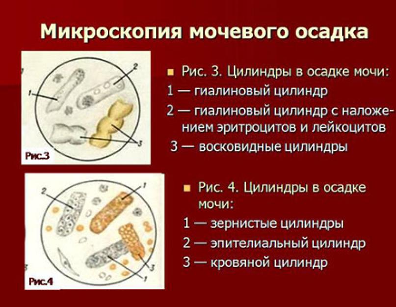
Cylinders in urine
Cylinders are elements of a cylindrical sediment (a kind of casts of renal tubules), consisting of protein or cells, may also contain various inclusions (hemoglobin, bilirubin, pigments, sulfonamides). According to the composition and appearance, several types of cylinders are distinguished (hyaline, granular, erythrocytic, waxy, etc.).
Normally, the cells of the renal epithelium secrete the so-called Tamm-Horsfall protein (absent in the blood plasma), which is the basis of hyaline casts. Hyaline casts can be found in the urine in all kidney diseases. Sometimes hyaline casts can be found in healthy people. As a pathological symptom, they acquire importance when constantly detected and in significant quantities, especially when erythrocytes and renal epithelium are superimposed on them.
Granular cylinders are formed as a result of destruction of tubular epithelial cells. Finding them in a patient at rest and without fever is indicative of renal disease.
Wax cylinders formed from compacted hyaline and granular cylinders in tubules with a wide lumen. They are found in severe kidney disease with a predominant lesion and degeneration of the epithelium of the tubules, more often in chronic than in acute processes.
Erythrocyte casts are formed when erythrocytes are layered on the hyaline casts, leukocyte - leukocytes. The presence of erythrocyte casts confirms the renal origin of hematuria.
Epithelial casts (rarely) are formed when tubular epithelium is detached. They occur with severe degenerative changes in the tubules at the beginning of acute diffuse glomerulonephritis, chronic glomerulonephritis. Their presence in the analysis of urine a few days after the operation is a sign of rejection of the transplanted kidney.
Pigment (hemoglobin) cylinders are formed when pigments are included in the cylinder, and is observed with myoglobinuria and hemoglobinuria.
Cylindroids - long formations of mucus. Single cylindroids are found in urine at normal levels. A significant number of them occur in inflammatory processes of the urinary tract mucosa. They are often observed when the nephritic process subsides.
Norm: hyaline cylinders - single, the rest - absent
Hyaline casts in urine:
- renal pathology (acute and chronic glomerulonephritis, pyelonephritis, kidney stones, renal tuberculosis, tumors);
- congestive heart failure;
- hyperthermic conditions;
- high blood pressure;
- taking diuretics.
Granular cylinders (nonspecific pathological symptom):
- glomerulonephritis, pyelonephritis;
- diabetic nephropathy;
- viral infections;
- lead poisoning;
- fever.
Wax cylinders:
- amyloidosis of the kidneys;
- nephrotic syndrome.
Erythrocyte casts (hematuria of renal origin):
- acute glomerulonephritis;
- kidney infarction;
- renal vein thrombosis;
- malignant hypertension.
Leukocyte casts (leukocyturia of renal origin):
- pyelonephritis;
- lupus nephritis with systemic lupus erythematosus.
Epithelial casts (most rare):
- acute tubular necrosis;
- viral infection (eg, cytomegalovirus);
- poisoning with salts of heavy metals, ethylene glycol;
- overdose of salicylates;
- amyloidosis;
- kidney transplant rejection reaction.
Bacteria in urine
Excretion of bacteria in the urine has significant diagnostic value. Bacteria persist in the urine for no more than 1-2 days after the start of antibiotic therapy. The first morning urine sample is preferred for research. It is possible to determine the type of bacteria and assess the level of bacteriuria, as well as to identify the sensitivity of microorganisms to antibiotics using bacteriological culture of urine.
Norm: negatively
Bacteria in the urine:
- infections of the urinary system, pyelonephritis, urethritis, cystitis).
Yeast fungi
- candidiasis, which occurs most often as a result of inappropriate antibiotic therapy.
Reference values: negatively
Inorganic urine sediment (crystals), salts in urine
Urine is a solution of various salts that can precipitate (form crystals) when urine stands. Low temperature favors crystal formation. The presence of certain crystals of salts in the urinary sediment indicates a change in the reaction to the acidic or alkaline side. Excessive salt content in urine contributes to the formation of calculi and the development of urolithiasis. At the same time, the diagnostic value of the presence of salt crystals in the urine is usually small. Increased doses of ampicillin and sulfonamides lead to the formation of crystals.
Uric acid under physiological conditions is found with a high concentration of urine, after plentiful meat food, after profuse sweating.
Norm: absent
Uric acid and its salts (urates):
- highly concentrated urine;
- acid reaction of urine (after exercise, meat diet, fever, leukemia);
- uric acid diathesis, gout;
- chronic renal failure;
- acute and chronic nephritis;
- dehydration (vomiting, diarrhea);
- in newborns.
Hippuric acid crystals:
- eating fruits containing benzoic acid (blueberries, lingonberries);
- diabetes;
- liver disease;
- putrefactive processes in the intestines.
Ammonia-magnesia phosphates, amorphous phosphates:
- alkaline urine reaction in healthy people;
- vomiting, gastric lavage;
- cystitis;
- fanconi's syndrome, hyperparathyroidism.
Calcium oxalate (oxaluria occurs with any urine reaction):
- eating foods rich in oxalic acid (spinach, sorrel, tomatoes, asparagus, rhubarb);
- pyelonephritis;
- diabetes;
- ethylene glycol poisoning.
Leucine and Tyrosine:
- severe metabolic disorder;
- phosphorus poisoning;
- destructive liver diseases;
- pernicious anemia;
- leukemia.
Cystine:
- congenital disorder of cystine metabolism - cystinosis.
Fat and fatty acids:
- degenerative changes in the epithelium of the renal tubules;
- eating a lot of fish oil.
Cholesterol:
- hiluria;
- fatty degeneration of the kidneys;
- echinococcosis of the kidneys;
- cystitis;
- cholesterol stones.
Bilirubin:
- hepatitis;
- liver cancer;
- infectious diseases;
- phosphorus poisoning.
Hematoidin:
- chronic bleeding along the urinary tract, especially if the blood stagnates anywhere.
Mucus in urine
Mucus is secreted by the epithelium of the mucous membranes. Normally present in urine in small quantities. In inflammatory processes, the content of mucus in the urine increases. An increased amount of mucus in the urine may indicate a violation of the rules for proper preparation for taking a urine sample.
Norm: insignificant amount.
When diagnosing pathological conditions of various kinds, a general urine analysis is mandatory. Urine sediment microscopy is part of the general clinical examination. The essence of this analysis is to calculate and assess the quality of the elements of the urinary sediment in order to identify or refute most pathological conditions.
Overview of the study
Correctly collected and delivered on time (delivery time 1-2 hours) to the laboratory material affects the correct interpretation of the analysis. For the analysis, you need a morning single portion of urine, stored in a sterile container for analysis. Be sure to perform intimate hygiene before starting the collection of the analysis.
Analysis of the indicator for suspicions of such pathological conditions:
- hematuria (erythrocyte count);
- pyuria (white blood cell count);
- cylindruria (cylinder counting);
- bacteriuria (number of bacteria)
- hemoglobinuria. The presence of salt crystals, epithelial cells, mucus, protozoa is also assessed.
 The study makes it possible to assess the function of the kidneys and urinary tract, as well as to identify violations in the work of internal organs.
 The research procedure consists of the following stages:
- With the help of a pipette, 10 ml of urine (standing for 1-2 hours) is collected from the bottom of the container.
- The collected material is centrifuged at 1500 rpm. lasting 5-7 minutes.
- The composition of one drop of sediment is analyzed using low and high magnification. The data obtained will help in diagnosing diseases, determining the ongoing inflammatory or infectious process, changes in metabolism.
Explanation of sediment analysis results: norm
It is important to bear in mind that some external factors can affect the result of the urine sediment microscopy analysis, such as:
- excessive physical activity;
- taking medications (diuretics or antibiotics);
- violation of the rules for collection and delivery of urine;
- diet.
The elements that are present in the sediment are usually classified as organized, having an organic origin, and unorganized - inorganic sediment. For all the identified elements of urinary sediment, there are certain norms, the excess of which indicates various pathological conditions.
Hemoglobin
 The presence of hemoglobin in the urine indicates severe infectious diseases.
 The presence of hemoglobin in the urine sediment is considered a deviation, since it is considered a sign of the breakdown of red blood cells. It is often caused by various diseases - flu, pneumonia, acute infection. But external reasons can also provoke its occurrence in urine, for example, hypothermia, injury, poisoning. In this case, the urine acquires a reddish-brown hue, painful sensations in the lumbar region may appear. Very often, urine saturated with hemoglobin indicates a failed blood transfusion when the donor's blood is incompatible with the patient's blood.
Erythrocytes in urine sediment
Erythrocytes in urine are unchanged and leached. Their difference lies in the content of hemoglobin, leached does not contain it. The leached ones present in the microscopic material indicate disorders in the functioning of the kidneys, unchanged ones - to violations of the urinary tract. The normal red blood cell count for the female sex is 3, for the male sex - 1. With other indicators, hematuria is noted. Allocate microhematuria (the color of urine does not change) and gross hematuria (urine changes color due to the presence of red blood cells). Avoid microscopic examination of urine sediment during critical days, as blood can enter the sample in the urine. The reasons for the appearance of hematuria include:
- kidney pathology and injury;
- diathesis;
- the presence of kidney stones;
- tumors of the urinary system;
- urinary tract infections;
- poisoning of various kinds.
Leukocytes in urine sediment
 A large number of white blood cells causes a cloudy urine.
 The boundary values \u200b\u200bof leukocytes in urine are 0-5 for women, 0-3 for men. If the values \u200b\u200bare increased, it means that pus is excreted in the urine (pyuria or leukocyturia). Pyuria always indicates an inflammatory focus. To determine the approximate localization of the passing inflammation, glass urine samples are prescribed, namely. The concentration of leukocytes in the first glass of urine is the initial pyuria, it indicates urethritis or prostatitis. End-pyuria is determined by the presence of leukocytes in the third glass and suggests cystitis. An excess of leukocytes in three glasses is ailments of the kidneys and bladder.
Epithelium in urine sediment
Epithelial cells (epithelial cells) are found in urine quite often, their presence in single values \u200b\u200bis considered the norm. Epithelial cells are classified by nature of origin. The renal epithelium identified in the sediment is of great importance, since this type of epithelium passes from the renal tubules. This fact indicates serious kidney damage. An increase in the permissible values \u200b\u200bof squamous epithelium often indicates an incorrect collection of analysis or an acute inflammatory process in the organs of the urinary system. Urine sediment containing polymorphic epithelium in values \u200b\u200bhigher than permissible carries information about diseases of the urinary tract (oncology, cystitis, stones, intoxication).
Microscopy of urine sediment is part of the laboratory study of clinical (general) urine analysis. The essence of the process lies in identifying or refuting various pathologies.
Microscopy of urine sediment is part of a general clinical medical examination of the human body to identify and diagnose a variety of pathological and chronic processes. The essence of studying the sedimentary environment of biological material is to assess the concentration and quality of the identified substances, which helps to obtain reliable results of health status.
Microscopic examination of urine sediment is part of a general urinalysis (OAM). A general urine analysis is able to identify a number of pathologies, chronic diseases, and diagnose the result of latent diseases. Ailments without specific symptoms of manifestation pose a serious threat, affecting many systems of vital activity over time.
General analysis of urine involves a laborious research process in laboratory conditions, part of which is reduced to the study of biomaterial sediment. The sediment is obtained by processing the liquid in a centrifuge, after which solid particles are detected at the bottom of the tube. From a glass container, substances (amorphous deposits, crystals and cells) suspended from urine are taken using a pipette onto a viewing glass under a microscope.
When examining the microscopy of a photo of sedimentary substances, it is permissible to see their display of schematic division. Micro sludge is divided into two types:
- Organized view.
The organized type of sediment contains organic matter, including epithelial cells, casts, erythrocytes and leukocytes.
- Unorganized type of connections.
This species includes inorganic elements: fungi, mucus, salts and bacteria.
The essence of the diagnostic technique, by studying the urine sediment, is to identify, review and collect information on the quantitative content of the following components:
- Leukocytes are cells circulating in the bloodstream, that's all the reasons for their presence in urine.
- Erythrocytes, the reasons for their presence in the urine are similar - microorganisms enter the biomaterial from the blood.
The reasons for the increased rate of red blood cells in urine, which a woman takes for analysis, may lie in the incorrect sampling of material during menstruation. It is recommended to postpone the analysis to other days, since particles getting into the biomaterial give a false positive result.
- The epithelium, which can be detected by microscopic examination of urine sediment, is renal, polymorphic and flat. The epithelium is the cellular tissue lining the surfaces of all mucous membranes and organ cavities without exception.
The presence of squamous epithelium in the sediment is considered normal. If the examination revealed the renal epithelium and polymorphic, then it is worth looking for the reasons that will reveal pathologies and various diseases of the body.
- Cylinders are protein coagulated compounds or cellular tissue of the renal epithelium. Substances are epithelial, leukocytic, erythrocytic, hyaline, waxy and granular.
The presence of hyaline cells of the cylinders in a single amount in the sediment is the norm. If the study found other types of cylinders, then the specialist begins to look for the reasons for the failure of the body's systems and pathological processes.
- Salts are also an integral part of the human body, and have many varieties: oxalates, urates, phosphates and others.
- Mucus is also found in the sediment. The reasons for the content of mucus in urine are simple - the substance is secreted by the epithelium of the mucous membrane of all organs.
- Bacteria should not be found in the composition. The reasons for their presence can be different. Depending on what causes infectious inflammation, a specialist prescribes an additional examination (bacteriological culture of urine) and appropriate adequate treatment.
In a laboratory study of urine sediment, a laboratory assistant, in order to avoid erroneous statement of results, uses a special atlas, where microscopy of urine sediment is decomposed. After all, for each substance being searched for, there is a special generally accepted content norm.
Results of the study of urine sedimentary material: norms
Interpreting the results is the specialization of the attending physician, who focuses on the performance of each patient individually:
- age;
- gender (male \\\\ female);
- level of physical activity;
- field of activity;
- way of life;
- general health.
Decoding the analysis of urine sediment microscopy is a diagnosis of the general state of human health. In the process, they rely on generally accepted norms for the presence of certain substances in a certain concentration:
- Leukocytes: in women less than 5, in women during pregnancy the same indicator, in men less than 3 in the field of view.
- Erythrocytes for both sexes and during pregnancy should be found no more than 2.
- Epithelium (flat) for women and during pregnancy ≤5, for men - ≤3.
- The renal epithelium should not be found. The likelihood of renal epithelial sediment during pregnancy is high and requires special attention.
- The transitional epithelium can be in the urine in a minimal amount.
- Hyaline cylinders are acceptable for detection. Other types of cylinders should be absent.
- Fungi and bacteria - complete absence. Fungi can be detected during pregnancy and in patients who are taking antibiotics.
- Mucus is acceptable in small amounts.
- Salts, although an integral part of the activity of many systems, should be absent in the sediment.
If any deviations from the norms of any indicator are found, the specialist should appoint additional examinations so as not to make an erroneous diagnosis.
Sediment in urine during pregnancy is especially carefully studied, since the slightest deviations, bacteria and other substances in the body can negatively affect the overall development of the child in the womb. During pregnancy, it is correct to take a urine test within the walls of the laboratory with the help of specialists in order to avoid the ingress of foreign microorganisms into the biological material. This fact is especially true during late pregnancy.
Microscopy of urine is an effective method of diagnosis, monitoring the course of the disease. A general urine test is also carried out in the case of screening examinations. In the clinical analysis of urine, the physicochemical parameters of urine and microscopy of urine sediment are assessed.
What affects the receipt of the result
The laboratory test result will not be reliable:
- In case of violation of the instructions for the delivery of the material (errors in hygiene procedures, sampling of material during menstruation).
- If the urine is stored for several hours before it reaches the laboratory.
- The presence of an excess amount of fluid entered the body before the test, non-compliance with dietary requirements.
- If parenteral administration of saline, glucose solutions, contrast agents were performed the day before.
- The presence of strong physical exertion, nervous disorders.
- In pregnant women.
- If the urethra is injured by a catheter or urethral smear.
- If the material is contaminated with hemorrhoidal blood.
- When taking medications that change individual indicators.
Urine collection
Preparing for urinalysis is preceded by intimate hygiene to prevent bacteria from entering. Take the morning urine obtained after waking up. The time allotted for the delivery of material to the laboratory is minimized. Collect urine in a sterile container, and then placed in a tube with a preservative.

The accuracy of the study depends on the purity of urine.
Physical indicators of urine
Microscopic examination of the physical parameters of urine includes: color, transparency, odor, urine density.
Colour
Urine contains pigments that give it a yellow color that differs in saturation. Color can change:
- diseases;
- food products;
- medical preparations.
Diseases accompanied by jaundice give the urine a dark, yellow tint. When blood gets in, a reddish tint appears. If there is an excessive amount of carrots in the diet, urine will turn orange, beets - reddish.
Clarity and smell
Normally, urine is transparent, after settling it becomes cloudy: salts precipitate. An increase in odor occurs when air is exposed to urine. With diabetes mellitus, a fruity shade is felt. From food they change the smell: onions, garlic, horseradish. Medicines have the same ability.
Density
At different times of the day, the density of urine is not the same. When the blood is filtered, the kidneys secrete primary urine. When the secretion is reabsorbed, it enters the bloodstream again. Secondary urine with a higher concentration is formed.
Chemical research
pH is the ratio of hydrogen ions to hydroxide ions. If their quantities are equal, it is a neutral solution. If imbalanced, the solution will be acidic or alkaline. Normal urine is characterized by a slightly acidic reaction. Shifts in the reaction of urine to one side or the other are dangerous with the formation of stones.
The shift to the acidic side occurs:
- if you consume an excessive amount of protein;
- with symptoms of fever;
- if the body is under heavy stress;
- with all types of fasting;
- if the body is affected by diabetes.
Alkaline:
- when rejecting proteins of animal origin;
- with frequent vomiting;
- with pathology of the urinary system.
Protein
Under normal conditions, there is no protein in the urine, or rather, it is in such an amount that it can be detected using special research methods, which is practically impractical.

Protein in urine indicates the presence of a disease
To establish the presence of protein, urine must be mixed with the desired reagent. The presence of protein is determined by the presence of a reaction: if a reaction has occurred, the protein is present in the urine. The presence of protein in urine is called proteinuria.
Functional proteinuria occurs in the following cases:
- if the body is exposed to physical activity;
- in case of nervous disorders;
- with the predominance of protein foods;
- with febrile symptoms.
Glucose
Normally, there should be no glucose in urine. Sometimes microscopic analysis notes a short-term appearance during glucose treatment or other increased absorption of sugar by the body. Most often, glucose is found in diabetes mellitus.
The presence of glucose in urine is called glucosuria. There are methods for detecting glucosuria. For this purpose, test systems are used - strips for applying urine droplets to monitor the color change. By comparing the changes with the rating scale, a conclusion can be made about the amount of glucose in the urine.
Ketone bodies
The detection of ketone bodies in urine is considered a deviation from the norm, the reason for this phenomenon is disturbances in the metabolism of fats and carbohydrates. If the body has to obtain energy from fat stores, when fat is broken down, ketone bodies are formed, which are excreted from the body along with urine. In this case, ketonuria takes place.

Ketone bodies in urine indicate a violation of the metabolism of carbohydrates and fats
The onset of ketonuria is facilitated by:
- strong physical activity;
- nervous disorders;
- diseases accompanied by fever;
- a diet that excludes carbohydrates;
- repeated vomiting.
Ketonuria is determined using a test strip with applied drops of urine, similar to the detection of glucosuria.
Bile pigments
Bilirubin, urobilin, bile acids at normal levels should not be present in the urine. If they appear, it means that the level of bilirubin in the blood is increased. In this case, there is the appearance of bound bilirubin, which is characteristic of hepatitis and obstructive jaundice.
Microscopy of urinary sediment
It takes about 2 hours to obtain urine precipitation. The elements in the sediment are divided into organized and unorganized. The former are of organic origin, the latter are inorganic. When microscopy urine, there are permissible norms for the presence of elements of urinary sediment. If these indicators are exceeded, we can assume the development of various pathologies.
Hemoglobin
When examining urine, hemoglobin is found in urine with severe infections of the body. Hemoglobin appears in urine during the breakdown of red blood cells. The causes of this condition are often diseases caused by various infections.
The appearance of hemoglobin in urine can be provoked by:
- repeated hypothermia;
- physical exercise;
- severe poisoning;
- physical exercise.
In such cases, the urine changes color, becomes reddish-brown, pain is felt in the lumbar region. Often, the urine is saturated with hemoglobin during unsuccessful blood transfusions. This occurs when the blood of the donor and the receiving patient is incompatible.
Erythrocytes
There are 2 types of red blood cells present in urine: unchanged and leached. The latter does not contain hemoglobin. Leached erythrocytes indicate kidney pathologies, unchanged ones - about disorders in the urinary system.
The norm of erythrocytes for a woman is 3, for a man - 1. If the indicators differ, there is hematuria, which is of two types: microhematuria, in which the color of urine remains unchanged, and gross hematuria (the color of urine changes due to the presence of red blood cells).
Hematuria (increased presence of red blood cells) appears in the following cases:
- with pathologies and injuries of the kidneys;
- with various forms of diathesis;
- if kidney stones are present;
- with oncological diseases of the urinary system;
- when toxic substances enter the body.
A lot of leukocytes in urine leads to clouding. For women, normal leukocyte counts in urine are considered 0-5, for men - 0-3. An increase in indicators indicates the presence of pus in urine, which signals the existence of inflammatory processes.

Leukocytes - cells of the human immune system
To determine where the inflammatory focus is, a glass urine sample is performed, in this case a three-glass sample. The first glass contains samples of initial pyuria, indicating the presence of urethritis, prostatitis. The number of leukocytes in 3 glasses determines the final pyuria, which often indicates a disease with cystitis.
An excess of the norm of leukocytes in three glasses indicates diseases of the urinary system.
Epithelium
The presence in urine is considered normal. An increase in the permissible value of squamous epithelium confirms severe inflammatory pathologies. Above normal polymorphic epithelium occurs in cancer, the presence of stones, lesions of various kinds of intoxication.
Cylinders
Found in various kidney diseases. The presence of hyaline cylinders is allowed only in a single quantity, since physical exertion is the reasons for the appearance. Other types of cylinders should normally be absent. Their presence in urine is called cylindruria.
The causes of cylindruria are:
- kidney disease;
- circulatory pathology;
- diseases accompanied by fever;
- getting into the body of various infections.
Unorganized sludge
Bacteria in urine
Normally, bacteria are not present in urine. Clinical analysis allows you to establish the presence of bacteria. To determine their type, bacteriological inoculation is carried out. If isolated bacteria are found, a diagnosis is made: bacteriuria. It is caused by urinary tract infections.
Quantitative study of urine
When using these methods, more accurate information is obtained regarding the elements included in the urinary sediment, and assistance is provided in the diagnosis of latent pyuria. Quantitative research with microscopy makes it possible to count the number of elements at a specific time in a specific volume.
It should be noted that in children, if indicated, urine samples are examined in the same way as in adults. Decoding of the results is carried out taking into account age norms. Often, to conduct a study of urinary sediment, the Nechiporenko technique is used, in which manual digital processing is replaced by fast and accurate counting according to a given execution algorithm.

The Nechiporenko technique is an effective way to study urine
The sequence of microscopy of urine according to Nechiporenko:
- The prepared urine is transferred into a centrifuge tube.
- Urine is centrifugally processed at a speed of 1500-2000 rpm for a quarter of an hour.
- The urine is separated from the sediment so that 1 ml of sedimentary urine remains in the test tube.
- A slide plate is taken and its chamber is filled with a drop of urine with sediment.
- The slide plate is placed on the microscope stage, and with its help the number of shaped elements is counted.
- Using a special formula, their amount is set in 1 ml of urine.
After obtaining the results for the diagnostic conclusion, use the atlas of microscopy of urinary sediment.


